¡Seguimos cuidando tu salud! Recuerda: el uso de cubrebocas es obligatorio durante tu estancia en el hospital; con esto evitamos la propagación de enfermedades respiratorias.

¡Seguimos cuidando tu salud! Recuerda: el uso de cubrebocas es obligatorio durante tu estancia en el hospital; con esto evitamos la propagación de enfermedades respiratorias.
Get an accurate and professional diagnosis, always with the warmth and efficiency that are with us in every service.
It is a diagnostic method that uses high frequency sound waves for imaging various organs and tissues.
When the sound beam passes through the human body it generates an interface between tissues of different densities, some of that energy is reflected and some is transmitted. The reflected waves are detected by the sensor or translator, and the transmitted power provides an image of scanned object.
The study is a simple, fast, economical and with a high degree of diagnostic certainty that there is no risk in pregnancy and when in doubt, is complemented by other imaging techniques such as radiology, computed tomography or MRI.
4D Ultrasound represents the moving image combined with volumetric reconstruction, in other words the surface structures. While the 3D technology it’s a reconstruction of the image through the computer in a static way.
There are multiple benefits offered in submitting to this procedure. For example, in gynecology, it determines the exact measurement of the volume of hyperplasia (endometrial), cysts, polyps, fibroids or myomas and keeps track of gynecologic tumor after treatment or chemotherapy and helps to evaluate the fallopian tubes confirming or not, sterility from this cause.
In obstetrics, detects abnormalities of the face, limbs, spinal canal, determines the exact position of the fetus, location of the placenta and umbilical cord and amniotic fluid characteristics. In smaller parts, serves as a guide for accurate biopsy, allowing accurate visualization of the needle and functions in evaluating the volume of a breast tumor. In urological terms aids in the visualization and correction of the position of a urinary catheter and measures the amount of residual urine.
In regards to internal medicine, facilitates the liver and kidney biopsies and helps to diagnose appendicitis, allows the precise localization of stones in the kidneys and has the ability to calculate the volume of the heart.
Ultrasonography can be used to study abdominal solid organs like liver, kidneys, spleen, pancreas, etc., or those containing liquid inside including: bladder, gall bladder, etc. It is also useful for exploring superficial organs such as muscles, tendons, mammary gland, scrotum, thyroid and pelvic cavities (male and female), and in some cases the hollow viscera (cecal appendix and stomach in infants).
It is used in special cases with endocavitary transducers, which allow the uterus or prostate cancer to be examined with higher resolution (transvaginal and transrectal ultrasound), and for the visualization of internal organs in endoscopic, intravascular examinations or during surgery.
We have the best equipment in high resolution. This service is available at the Unit of Radiology and Imaging in Médica Sur Tlalpan and Medica Sur Lomas.
El ultrasonido es un método de diagnóstico no invasivo e indoloro, no utiliza radiación y que usa ondas sonoras de alta frecuencia para la formación de imágenes de los distintos órganos, tejidos corporales y estructuras internas que ayudarán a tu médico a conocer:
Es uno de los estudios más seguros, económicos e importantes que no produce radiación ionizante y pueden durar de 30 minutos a 1 hora con un alto índice de certeza diagnóstica.
El ultrasonido de diagnóstico examina de forma fácil y útil muchos órganos internos como el corazón, vasos sanguíneos, ojos, tiroides, cerebro, tórax, órganos abdominales, piel, músculos y muchos más. Por ello, tiene un gran número de aplicaciones en diferentes especialidades y áreas médicas, por ejemplo:
La preparación para tu estudio dependerá del tipo de examen que te hagan. En algunos casos puede ser necesario que no comas ni bebas nada durante 12 horas anteriores al examen, o bien, necesitarás tomar seis vasos de agua dos horas antes del estudio, sin orinar, para visualizar mejor los órganos. Siempre pregunta a tu médico las condiciones que requieres y resuelve tus dudas.
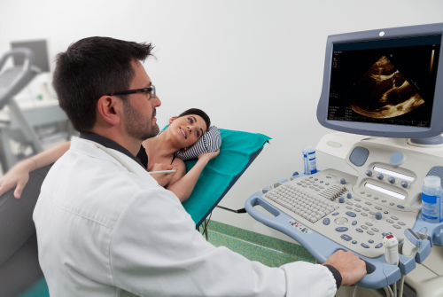
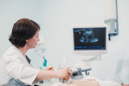
Para este estudio no se necesita preparación alguna, solo usar ropa cómoda y que ayude a descubrir fácilmente el abdomen.
Evalúa el sistema circulatorio del cuerpo para conocer:
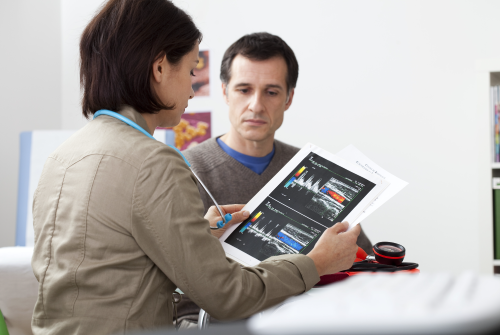
Así como guiar la colocación de agujas o catéter en venas o arterias.
Este estudio no requiere preparación especial, en algunos casos puedes necesitar ayuno.
Es empleado como una herramienta diagnóstica de los tejidos blandos, nervios, músculos, tendones y ligamentos de hombro, codo, muñecas, manos, dedos, cadera, rodilla, tobillo y pie; gracias a su detalle anatómico y visualización en varios planos para conocer enfermedades o patologías como:
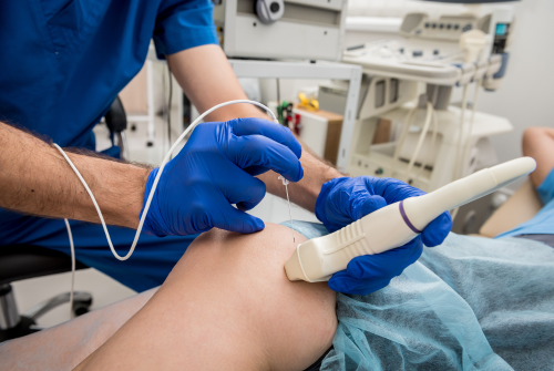
Estos estudios no requieren preparación especial.
En Médica Sur contamos con los mejores equipos tecnológicos de alta resolución para darte un diagnóstico certero. Contáctanos y resuelve las dudas del estudio que necesitas en la Unidad de Radiología e Imagenología.
¡Pregunta por tu estudio y agenda cita! 55 5424 7200 ext. 7215, 7216
Información sujeta a cambio sin previo aviso 11/AGO/2025 LFAL
Do you live outside Mexico?
We give you advice and support for your travel and stay al Médica Sur
Toll free: 800.501.0101
Canada / USA: 1877.213.6659
24 hours everyday