¡Seguimos cuidando tu salud! Recuerda: el uso de cubrebocas es obligatorio durante tu estancia en el hospital; con esto evitamos la propagación de enfermedades respiratorias.
¡Seguimos cuidando tu salud! Recuerda: el uso de cubrebocas es obligatorio durante tu estancia en el hospital; con esto evitamos la propagación de enfermedades respiratorias.
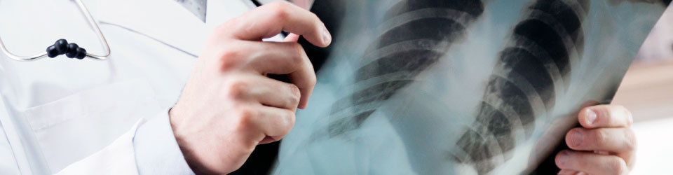
Studies the anatomical regions of the orbits and their contents (eyes, muscles and nerves); middle and inner ear, with its different components, nose and sinuses, throat (larynx, pharynx, glands and vascular structures), maxilla and mandible. All these studied by computed tomography.
The advantages of CT are: possibility shortening cuts to less than 1 mm due to its resolution of .33 mm; appreciates different types of tissues such as bone, fat, muscle, glandular tissue, and so on; uses intravenous contrast medium to assess the vascular behavior of the pathology to be evaluated.
The main benefit is that thanks to the advantages mentioned above, it is possible to evaluate the structures of interest in multiple planes and three-dimensionally, allowing us an accurate diagnosis.
Specifically in the temporal bone and cerebellopontine angle, it is possible to evaluate the middle and inner ear and the inner and outer ear canal as well as the study of infectious or inflammatory diseases, tumors, vascular disease and congenital conditions that can cause hearing loss.
In vascular structures, allows for non-invasive diagnostic vascular lesions (arteries or veins), especially in the neck, using the technique of angiography.
For dental cases, we use special software that allows examination for tumors of this region, infectious and or inflammatory diseases, as well as planning for implant surgery.
In orbits, it is useful for the evaluation of tumors of the muscles or the optic nerve and the eyeball, also for the evaluation of pseudo causing alterations in the size and shape of the eyes.
For nose and sinuses, is used for evaluation, diagnosis and flattening of fractures, tumors, infections and inflammations.
In the neck, is it possible to assess the upper airway (pharynx and larynx) and lymph node and muscle diseases.
Información sujeta a cambio sin previo aviso 11/AGO/2025 LFAL
Do you live outside Mexico?
We give you advice and support for your travel and stay al Médica Sur
Toll free: 800.501.0101
Canada / USA: 1877.213.6659
24 hours everyday