¡Seguimos cuidando tu salud! Recuerda: el uso de cubrebocas es obligatorio durante tu estancia en el hospital; con esto evitamos la propagación de enfermedades respiratorias.
¡Seguimos cuidando tu salud! Recuerda: el uso de cubrebocas es obligatorio durante tu estancia en el hospital; con esto evitamos la propagación de enfermedades respiratorias.
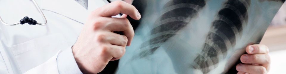
A medical examination that is noninvasive and painless that obtains special X-ray images to produce multiple views inside the body. Then, the images can be printed or reviewed on a computer monitor or workstation.
CT scans of internal organs, bones, soft tissues and blood vessels provide greater clarity than conventional x-ray examinations
The advantages and benefits of using this technology are the quickness of the procedure for patients with lesions on the chest, because internal injury or bleeding can be diagnosed on time and be offered a treatment that can save a life. In addition, CT images are accurate, noninvasive and painless.
It also provides real-time imaging, making it a good tool to guide procedures such as biopsies of lungs, abdomen, pelvis and bones.
In general, CT imaging is not recommended for pregnant women because of potential risk to the baby. Also, breastfeeding mothers should wait 24 hours after receiving the IV injection of contrast material before resuming breast-feeding.
As for children, the study can be applied to them only if it is essential for making a diagnosis and should not be performed repeatedly in unless absolutely necessary.
Diagnosis: The chest CT scan is used to further examine and diagnose abnormal chest signs of disease, to evaluate whether tumors are responding to treatment, helps to program radiation therapy, helps to detect vascular occlusion, and detect thoracic malformations.
A CT scan of the chest can also be used to explore lung cancer in patients that are smokers or former smokers who have a much higher risk of getting cancer than nonsmokers.
The chest CT scan may also show lung conditions such as past or current pneumonia, tuberculosis, emphysema, bronchiectasis, inflammation and interstitial disease.
A CT scan angiogram may be performed to evaluate blood vessels (arteries and veins) in the chest. It involves injecting iodine into the vein a little faster and also getting a larger number of more detailed image slices through the chest to see the arteries in the best possible way.
Información sujeta a cambio sin previo aviso 11/AGO/2025 LFAL
Do you live outside Mexico?
We give you advice and support for your travel and stay al Médica Sur
Toll free: 800.501.0101
Canada / USA: 1877.213.6659
24 hours everyday